
What Is the Function of Primers in a PCR Reaction?
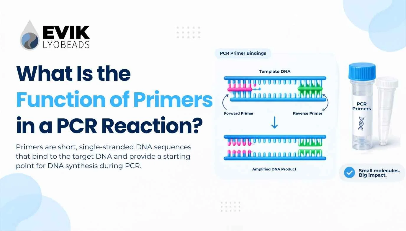
Ask what is the function of primers in a PCR reaction and the textbook answer comes back immediately: primers tell the polymerase where to start. True, and not nearly enough. That answer explains why you need primers but not why a badly designed pair will cost you three weeks, why the same primers behave differently at 58 °C and 62 °C, or why nearly every commercial master mix on the market deliberately leaves them out.
Primers are two short synthetic oligonucleotides, usually 18 to 30 bases each, and they carry more of the reaction's behaviour than any other component. The polymerase is generic. The buffer is generic. The dNTPs are generic. The primers are the only part of a PCR that knows what you are looking for.
This article covers the chemistry that makes primers mandatory, the five distinct jobs a primer pair performs, what separates a good primer from one that will waste your reagents, and why primers sit outside the master mix in almost every workflow.
The Short Answer
In a PCR reaction, primers perform five functions at once:
They provide the free 3′-hydroxyl group that DNA polymerase chemically requires to add the first nucleotide. Without one, no synthesis occurs at all.
They define the amplicon. The forward primer marks one boundary, the reverse primer marks the other, and everything between them is copied.
They supply specificity. Out of a three-billion-base human genome, the primer pair selects one region.
They set the annealing temperature. The primers' melting temperature dictates a key thermal parameter of the entire cycling programme.
They make amplification exponential. Two opposing primers, rather than one, are what turn linear copying into doubling.
Each of these deserves unpacking, because each one is a place where a reaction fails.
Why PCR Cannot Start Without a Primer
This is the part usually skipped, and it is the reason primers exist.
DNA polymerase does not build a strand from nothing. It extends an existing one. The catalytic step is a nucleophilic attack by a free 3′-hydroxyl group on the alpha-phosphate of the incoming deoxynucleotide triphosphate, which forms the phosphodiester bond and releases pyrophosphate. That reaction needs a 3′-OH already in place, correctly positioned and base-paired to the template.
No known DNA polymerase can initiate synthesis de novo. This is a hard constraint on the enzyme class, not a limitation of Taq specifically. It is exactly why living cells use primase — a specialised RNA polymerase — to lay down short RNA primers during chromosome replication. RNA polymerases can start from a naked single strand; DNA polymerases cannot.
PCR uses synthetic DNA oligonucleotides instead of RNA primers, but the requirement is identical. The primer anneals to the template, presents its 3′ end as a starting point, and the polymerase takes over from there.
One practical consequence follows directly: the 3′ end of a primer is the business end. A mismatch near the 5′ end is often tolerated and the reaction proceeds. A mismatch in the last two or three bases at the 3′ end can kill amplification outright, because the polymerase cannot properly seat a poorly paired 3′ terminus. Every primer design rule about 3′ ends traces back to this single fact.
The Five Jobs, in Detail
1. Providing the Chemical Starting Point
Covered above, and worth restating in its practical form: primers are consumed by the reaction. They are not catalysts. Each primer molecule that anneals and extends becomes physically incorporated into a product strand, permanently. This is why primer concentration matters and why primer depletion contributes to the plateau phase in qPCR — late in a reaction, primers and dNTPs run short while the accumulating product competes for them.
Typical working concentration is 0.1–0.5 µM per primer, with a usable range of roughly 0.05–1 µM. Higher is not better. Excess primer promotes non-specific annealing and primer-dimer formation, both of which consume reagents while producing nothing you want.
2. Defining the Amplicon
The forward primer binds the antisense strand and points inward. The reverse primer binds the sense strand and points inward from the other side. Synthesis proceeds 5′ to 3′ from each, toward the other.
The region between the two 5′ ends is your amplicon, and its length is fixed entirely by where you place the primers. Nothing outside that window gets amplified.
This has design consequences beyond the obvious. For qPCR, short amplicons of 80–150 bp are standard because they amplify with higher efficiency and tolerate partially degraded template. For applications requiring the amplicon to be sequenced or cloned, longer products are necessary and primer placement changes accordingly. If you need to distinguish cDNA from genomic DNA in an RT-PCR assay, you place primers across an exon–exon junction so that genomic template will not amplify. All of this is primer placement, decided before a single reagent is pipetted.
3. Delivering Specificity
A 20-base sequence has 4²⁰ possible variants, roughly one trillion. Statistically, a specific 20-mer should occur once in a genome of three billion bases. That is the theoretical basis for primer specificity.
Practice is less tidy. Primers tolerate mismatches, especially away from the 3′ end. Genomes contain repeats, pseudogenes, and paralogous families. A primer that looks unique in isolation may have dozens of near-matches in real template. This is why in silico specificity checking with Primer-BLAST is not an optional refinement — it is the step that catches the mispriming you would otherwise discover at the gel.
Specificity is also thermal, not just sequence-based, which leads directly to the next function.
4. Setting the Annealing Temperature
The primers' melting temperature (Tm) is the temperature at which half the primer–template duplexes have dissociated. The annealing step of your cycling programme is set relative to it, conventionally around 5 °C below the lower primer Tm, and then optimised empirically.
This creates the specificity–yield trade-off that dominates PCR optimisation:
Annealing too low — primers bind at partially matched sites as well as perfect ones. You get product, plus non-specific bands, plus dimers.
Annealing too high — primers barely bind even correct sites. You get clean results and very little of them.
Because both primers must work at the same annealing temperature, their Tm values need to be close. Most guidelines specify within 5 °C, and within 2 °C is better. A pair with Tm values of 55 °C and 68 °C has no annealing temperature that suits both, and no amount of optimisation fixes that. It is a design defect.
Note that Tm is not a fixed property of the sequence alone. Salt concentration, magnesium, and primer concentration all shift it, which is why a primer pair validated in one buffer system can behave differently in another. Reformulating an assay — including drying it down into a stabilised format — is a case where re-verifying annealing behaviour is worth the effort rather than assumed.
5. Making the Reaction Exponential
This function is the easiest to overlook and arguably the most important.
With a single primer, each cycle produces one new copy per template strand. Copies accumulate linearly: after 30 cycles you have roughly 30 copies. Useless for detection.
With two opposing primers, each new strand contains the binding site for the other primer. Products become templates. Copy number doubles each cycle rather than incrementing: 2ⁿ, which after 30 cycles is over a billion-fold amplification.
That inward-facing geometry is what makes PCR work. It is not a convenience. Remove one primer and you have linear amplification, which is a genuinely different technique with different uses.
How Primers Behave Across the Three Steps of a Cycle
Denaturation (94–98 °C). Double-stranded template separates. Primers are single-stranded and unbound, floating free. Nothing anneals at this temperature.
Annealing (50–65 °C, typically). Temperature drops and primers bind their complementary sites. Primers win this race against template re-annealing for two reasons: they are present in vast molar excess, and being short, they diffuse and hybridise far faster than two long genomic strands can find each other. This kinetic advantage is the reason the whole method functions.
Extension (68–72 °C). Polymerase binds the primer–template junction and synthesises the new strand from each primer's 3′ end. Taq extends at roughly 1,000 bases per minute at 72 °C, which is where the standard rule of one minute per kilobase comes from.
Forward and Reverse Primers: What Actually Differs
The two primers are not chemically different. Both are single-stranded DNA oligos with a free 3′-OH. The distinction is purely positional.
The forward primer matches the sense strand sequence and anneals to the antisense strand. The reverse primer is the reverse complement of the sense strand at the downstream boundary and anneals to the sense strand.
The most common design error among people new to PCR is writing the reverse primer as the sequence read from the reference directly, rather than its reverse complement. The result is a primer that binds nothing. Modern design software handles this automatically, which is one of several good reasons not to design primers by hand.
What Makes a Good Primer
ParameterTargetReasonLength18–30 nt (20–25 ideal)Long enough to be unique, short enough to anneal efficientlyTm55–65 °CCompatible with standard cycling; qPCR often targets ~60 °CTm difference within pair≤2–5 °CBoth must work at one annealing temperatureGC content40–60%Balanced binding strength; extremes cause secondary structure or weak bindingGC clamp1–2 G/C in final 3′ basesStabilises the 3′ end where the polymerase startsHomopolymer runs<4 identical consecutive basesRuns promote slippage and misalignment3′ complementarity≤3 contiguous bases3′ pairing between primers produces extendable dimersHairpin stabilityΔG less negative than about −5 kcal/molSelf-structure competes with template bindingSpecificityConfirmed by Primer-BLASTIn silico uniqueness before wet-lab work
The GC clamp deserves a note. One or two G/C bases at the 3′ terminus strengthen that end through three hydrogen bonds per pair instead of two, stabilising the exact position where polymerase initiates. But a long GC stretch there is counterproductive — it raises the risk of stable primer-dimer formation. One or two bases, not five.
When Primers Go Wrong
Most PCR failures are primer failures. The common modes:
Primer-dimers. Two primers anneal to each other, typically through complementary 3′ ends, and the polymerase extends them into a short double-stranded product. Because dimers are short, they amplify very efficiently and can consume the reaction. In SYBR-based qPCR they generate signal indistinguishable from real product at the detection step, which is why melt curve analysis is mandatory. Design tools screen for this: a maximum acceptable 3′ dimer ΔG of about −2.0 kcal/mol is a common threshold.
Hairpins. A primer with internal self-complementarity folds back on itself. The folded form cannot anneal to template, so effective primer concentration drops and yield falls.
Mispriming. Partial matches elsewhere in the template get extended, producing extra bands or, worse, a single clean band of the wrong sequence. Raising annealing temperature or using a hot-start polymerase usually resolves it.
Template secondary structure. GC-rich regions and stable hairpins in the template block primer access. Additives such as DMSO or betaine, or moving the primer site, are the usual remedies.
Degraded or poorly quantified primer stock. Repeated freeze-thaw cycles degrade oligos. Primers stored as working dilutions in water rather than TE degrade faster still. When a previously reliable assay drifts for no apparent reason, the primer stock is a leading suspect.
Primers in Formats Beyond Standard PCR
qPCR. Same primer function, tighter constraints. Amplicons of 80–150 bp, Tm near 60 °C, and stringent dimer screening because dimers generate false signal in intercalating-dye chemistries.
RT-qPCR. Primers must account for the RNA-to-cDNA step. Exon-spanning designs prevent genomic DNA contamination from producing signal. Assays of this type are the standard use case for RT-qPCR master mix formats, with the primer set supplied separately by the assay developer.
Probe-based assays. A TaqMan-style probe adds a third oligo binding between the primers, providing a second layer of specificity. The primers' function is unchanged; the probe reports rather than primes.
Multiplex PCR. Several primer pairs in one tube. Difficulty scales steeply, because every primer can potentially dimerise with every other primer. A four-plex has 28 possible pairwise interactions to screen.
Isothermal methods. LAMP takes primer function furthest, using four to six primers that recognise six to eight distinct regions of the target. The extra primers are what allow amplification without thermal cycling and what give LAMP its notable specificity — but they also make LAMP primer design substantially harder than PCR primer design. RT-LAMP master mix formats likewise leave the primer set to the developer.
Why Primers Are Left Out of the Master Mix
This question follows naturally from everything above, and the answer is a useful test of whether the concepts have landed.
A master mix contains the universal components: polymerase, dNTPs, buffer, magnesium, and stabilisers. Every PCR needs those, in essentially the same form, regardless of target. They can be manufactured in bulk, validated once, and used across every assay in a lab.
Primers are the opposite. They are the target-specific component. A mix containing primers is not a master mix — it is a single assay, useful for exactly one target and worthless for anything else.
There is a second, more technical reason. Primers stored in a mix alongside active polymerase have time and opportunity to form dimers, and any dimer that forms during storage is amplifiable the moment the reaction starts. Keeping primers separate until reaction setup removes that failure mode.
This is why lyophilized master mix beads are formulated with everything except primers, probes, and template. The user adds their own primer set at setup, which keeps one bead format compatible with any assay they develop. It also means the shelf-stable, cold-chain-free component is the part that is genuinely universal, while the part that changes between assays stays under the developer's control. Practical detail on how this works in a real workflow is covered in the qPCR master mix bead guide and the RT-LAMP bead guide.
Teams that eventually do want primers dried down into a complete single-tube assay face a real formulation question, since oligos have different stabilisation requirements from enzymes. Screening excipient compatibility across the full component set — enzyme, primers, probe, buffer — is what excipient selection plates are built for.
Frequently Asked Questions
What is the function of primers in a PCR reaction, in one sentence?
Primers give DNA polymerase a chemical starting point with a free 3′-OH, and by binding at two defined positions they select which region of the template gets amplified.
Why does PCR need two primers instead of one?
Two opposing primers make amplification exponential. Each new strand contains a binding site for the other primer, so products become templates and copy number doubles every cycle. One primer gives linear accumulation, which is far too slow to be useful for detection.
Are PCR primers DNA or RNA?
DNA. Cells use RNA primers made by primase during replication, but PCR uses synthetic single-stranded DNA oligonucleotides, which are more stable and are not removed afterward — they become part of the product.
How long should a PCR primer be?
18–30 nucleotides, with 20–25 optimal for most applications. Shorter primers lose specificity; longer ones anneal less efficiently and cost more to synthesise.
What happens if primer Tm values do not match?
There is no annealing temperature that works for both. The lower-Tm primer either binds non-specifically at a temperature that suits its partner, or fails to bind at all. Redesign is the fix, not optimisation.
Do primers get used up during PCR?
Yes. Primers are incorporated into the product strands and are consumed stoichiometrically. Their depletion is one contributor to the plateau phase seen at the end of a qPCR amplification curve.
Can I put primers in a lyophilized master mix?
It is possible but it converts a general-purpose reagent into a single-target one, and it requires separate formulation work because oligos and enzymes have different stabilisation needs. Most workflows keep primers separate for exactly this reason.
The Practical Takeaway
Returning to the question of what is the function of primers in a PCR reaction: they are not a supporting reagent. They are the specification of the assay. The polymerase, buffer, and nucleotides are interchangeable infrastructure. The primers determine what is amplified, how specifically, at what temperature, and whether the reaction produces clean product or a tube full of dimers.
Which is why primer design deserves more time than most workflows give it, and why the components around the primers should be as reliable and as invariant as possible. When the master mix is a known quantity, an assay that fails points somewhere useful.
EVIK™ DxSpheres are ready-to-use lyophilized reagent beads containing every component of a qPCR, RT-qPCR, or RT-LAMP reaction except primers, probes, and template — room-temperature stable, no cold chain, dissolving in seconds at setup. Browse the full product range, review the technical FAQs, or contact our team about custom formulations for your assay.